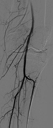
 An angiogram is a minimally invasive method that involves using an xray or “fluoroscopy” or “c-arm” machine, allowing your surgeon to visualize the arterial system, and the arteries in the abdomen, pelvis, and chest that would not be able to be seen with duplex scanning.
In general, arteriograms are performed when duplex scanning shows moderate to severe arterial disease to further define the anatomy in detail.
This is still the gold standard for imaging the arterial system of the trunk, abdomen, and pelvis, and to clearly delineate the extent of atherosclerosis or arterial narrowing. It is safely performed in an outpatient setting in our accredited operating facilities.
It involves the placement of a small IV catheter in the artery, through which dye is injected that outlines the arterial system making it “light up” under active xray imaging.
One of the major benefits of arteriography, is that it can be actively used for other procedures of the arterial system, without the need for a traditional incision. Instead, xray guidance and small, technologically advanced catheters are used to obtain the same result in most cases.